Home › Urethral Stricture Disease › 3D Anatomy Atlas
Urethral Stricture in 3D: An Interactive Anatomy Atlas
By Alex J. Vanni, MD, FACS — Director, Center for Reconstructive Urologic Surgery, Lahey Clinic
One urethra, one short stricture, and every way of treating it. Rotate the model, open it in cutaway to see the scar inside the channel, then step through each treatment to see what it does to the scar and why the results differ.
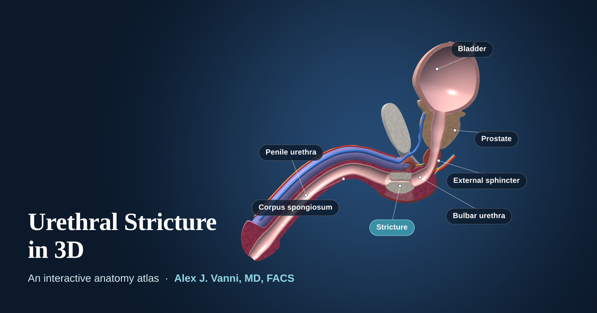
The interactive model needs WebGL, which is not available in this browser. The anatomy and each treatment are described in full below.
Schematic teaching model. Anatomy is simplified and not to scale in every detail. For education only, not for clinical use.
How to Use the Atlas
- Look aroundDrag to rotate. Pinch, or hold Ctrl and scroll, to zoom. Shift-drag moves the model.
- Look insideCutaway opens the model along the midline. X-ray view shows the channel the way a urethrogram does.
- Ask what something isClick any structure for its name and what it has to do with stricture disease.
What the Five Views Show
-
The stricture
A urethral stricture is scar tissue that narrows the channel urine passes through. The scar starts in the lining of the urethra and often extends into the spongy tissue around it, which is called spongiofibrosis. The model shows a 1.5 cm stricture in the bulbar urethra, the most common location, narrowing a channel about 10 mm wide to under 2 mm.
-
Dilation and internal urethrotomy (DVIU)
The scar is stretched with dilators, or cut from the inside with a small blade passed through a scope. Both open the channel, but neither removes the scar, and the cut heals by forming new scar. These procedures can provide short-term relief, but they carry high recurrence rates compared with urethroplasty, particularly for longer or recurrent strictures. More on why strictures recur.
-
Drug-coated balloon dilation (Optilume)
A balloon stretches the stricture open while its coating delivers paclitaxel, a medication that slows scar-forming cells, into the urethral wall. It is an option for selected men, typically those with a short bulbar stricture that has returned after earlier dilation or urethrotomy. Like those procedures, it leaves the scar in place. Watch: Optilume, the real-world story.
-
Excision and primary anastomosis
For a short bulbar stricture, the scarred segment can be removed completely and the two healthy ends sewn together in a wide, tension-free join. Because the scar is removed rather than stretched, this is a durable repair. In Dr. Vanni’s practice, five-year success for this repair is approximately 97%.
-
Buccal mucosa graft urethroplasty
For longer strictures, or when removing a segment would shorten the urethra too much, the urethra is opened lengthwise through the scar and a patch of buccal mucosa, the lining of the inner cheek, is sewn in to widen the channel. The graft can be placed on the top (dorsal) or bottom (ventral) of the urethra. The atlas shows both. In Dr. Vanni’s practice, five-year success is approximately 93 to 97%, depending on patient factors.
Urethroplasty is the gold-standard treatment for most urethral strictures and offers the highest chance of long-term cure. After either type of repair, a catheter rests in the urethra while it heals, typically for 1 to 3 weeks. See recovery after urethroplasty and the urethral stricture FAQs.
The Parts of the Male Urethra
- Prostatic urethra
- The first part, running through the prostate just below the bladder.
- Membranous urethra
- A short segment wrapped by the external sphincter, the muscle that provides urinary control.
- Bulbar urethra
- The widest part, deep in the perineum between the scrotum and the anus. The most common place for a stricture.
- Penile urethra
- Runs the length of the penis. Strictures here are usually repaired with a graft rather than by removing a segment.
- Fossa navicularis and meatus
- The last widening inside the head of the penis, and the opening at the tip. Often involved in lichen sclerosus.
About This Model
The atlas is a schematic built for teaching. Shapes and proportions are simplified, the stricture is a constructed example rather than a real patient, and no two strictures are alike. It is not a substitute for an individual evaluation, which usually includes a flow test, a retrograde urethrogram and often cystoscopy.
This page is for educational purposes and is not a substitute for an individual medical evaluation. Reviewed by Alex J. Vanni, MD, FACS. Last updated September 19, 2026.
Questions About a Urethral Stricture?
Dr. Vanni evaluates patients with primary and recurrent urethral strictures from across New England and nationwide.
Call (781) 744-8762 or learn more about urethral stricture treatment.