Home › Urethral Stricture Disease › Fossa Navicularis 3D Surgical Atlas
Fossa Navicularis Stricture in 3D: An Interactive Surgical Atlas
By Alex J. Vanni, MD, FACS — Director, Center for Reconstructive Urologic Surgery, Lahey Clinic
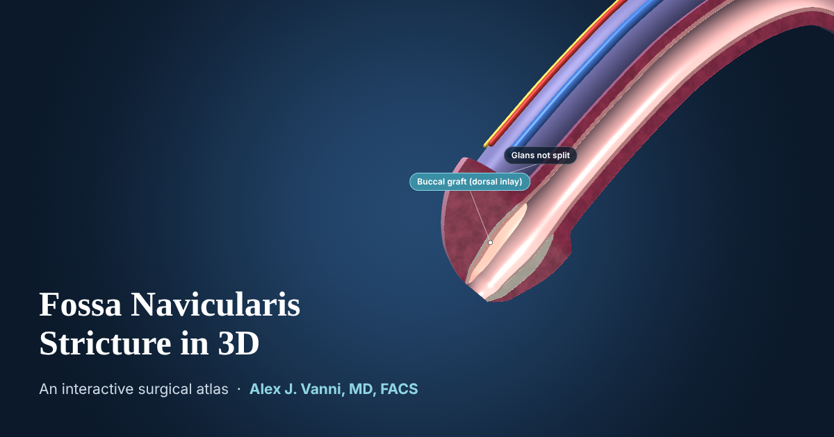
A stricture at the very tip of the urethra, and a repair done entirely through the opening. Rotate the model, open it in cutaway to see the scar inside the head of the penis, then follow the transurethral dorsal inlay buccal graft repair that Dr. Vanni described and published, which widens the channel without splitting the glans.
The interactive model needs WebGL, which is not available in this browser. The anatomy and each treatment are described in full below.
Schematic teaching model. Anatomy is simplified and not to scale in every detail. For education only, not for clinical use.
How to Use the Atlas
- Look aroundDrag to rotate. Pinch, or hold Ctrl and scroll, to zoom. Shift-drag moves the model.
- Look insideCutaway opens the model along the midline. Meatus view looks straight into the opening, the route the repair takes.
- Ask what something isClick any structure for its name and what it has to do with the stricture.
What the Two Views Show
-
The stricture
The fossa navicularis is the last part of the urethra, a small widening inside the head of the penis (the glans) just before the opening at the tip (the meatus). A stricture here is scar that narrows the outlet of the whole urinary tract, causing a weak or spraying stream, straining, burning with urination and incomplete emptying. Lichen sclerosus and previous transurethral surgery or catheterization are the most common causes. The model shows a 1.5 cm stricture extending back from just inside the meatus.
-
Transurethral dorsal inlay buccal graft urethroplasty
Working entirely through the meatus, the scarred roof (dorsal side) of the fossa navicularis is opened lengthwise, through the stricture and into healthy urethra beyond it. A graft of buccal mucosa, the lining of the inner cheek, is tailored to fit, drawn in through the opening and stitched into the incision with fine absorbable sutures, widening the channel. The glans is never split, so there is no glans incision to heal and its natural appearance is preserved. A catheter stays in place for about one week while the graft heals.
Results of the Repair
Dr. Vanni described this glans-sparing technique and published his results in the World Journal of Urology in 2022 (Farrell, Campbell, Zhang, Nowicki and Vanni). Approximately 94% of repairs were successful at a median follow-up of more than two years. Spraying of the urinary stream became less common after surgery, no man developed new spraying, and penile sensation was unchanged in most men.
Traditional open repairs of the fossa navicularis often require splitting the glans, which can affect its appearance and function. Avoiding that incision also removes the risk of the glans separating as it heals.
Other Options
Some men are first treated with dilation, with or without intermittent self-catheterization to keep the channel open, or with an extended meatotomy, which widens the opening at the tip. These can help, but the scar remains. For a stricture that returns, a reconstruction such as the transurethral dorsal inlay offers a more durable result. Dr. Vanni reviews every option with each patient. See the urethral stricture FAQs and recovery after urethroplasty.
Where the Fossa Navicularis Sits
- Penile urethra
- Runs the length of the penis beneath the erectile bodies and leads into the fossa navicularis.
- Fossa navicularis
- The last widening of the urethra, inside the glans. Lichen sclerosus often begins here and at the meatus.
- Meatus
- The opening at the tip of the penis. In the transurethral repair, all of the work is done through it.
- Glans
- The head of the penis, formed by the end of the corpus spongiosum. It surrounds the fossa navicularis.
For a stricture deeper in the urethra, see the Bulbar Urethral Stricture in 3D atlas.
About This Model
The atlas is a schematic built for teaching. Shapes and proportions are simplified, the stricture is a constructed example rather than a real patient, and no two strictures are alike. It is not a substitute for an individual evaluation, which usually includes a flow test, a retrograde urethrogram and often cystoscopy.
This page is for educational purposes and is not a substitute for an individual medical evaluation. Reviewed by Alex J. Vanni, MD, FACS. Last updated October 17, 2026.
Questions About a Fossa Navicularis Stricture?
Dr. Vanni evaluates patients with fossa navicularis, meatal and lichen sclerosus strictures from across New England and nationwide.
Call (781) 744-8762 or learn more about urethral stricture treatment.