Home › Ureteral Stricture › 3D Surgical Atlas
Ureteral Stricture in 3D: An Interactive Surgical Atlas
By Alex J. Vanni, MD, FACS — Director, Center for Reconstructive Urologic Surgery, Lahey Clinic
One ureter, and a repair matched to where the narrowing is and how long it runs. Rotate the model, switch to the X-ray view to see the narrowing and the swelling above it, then step through each reconstruction to see how the gap is bridged.
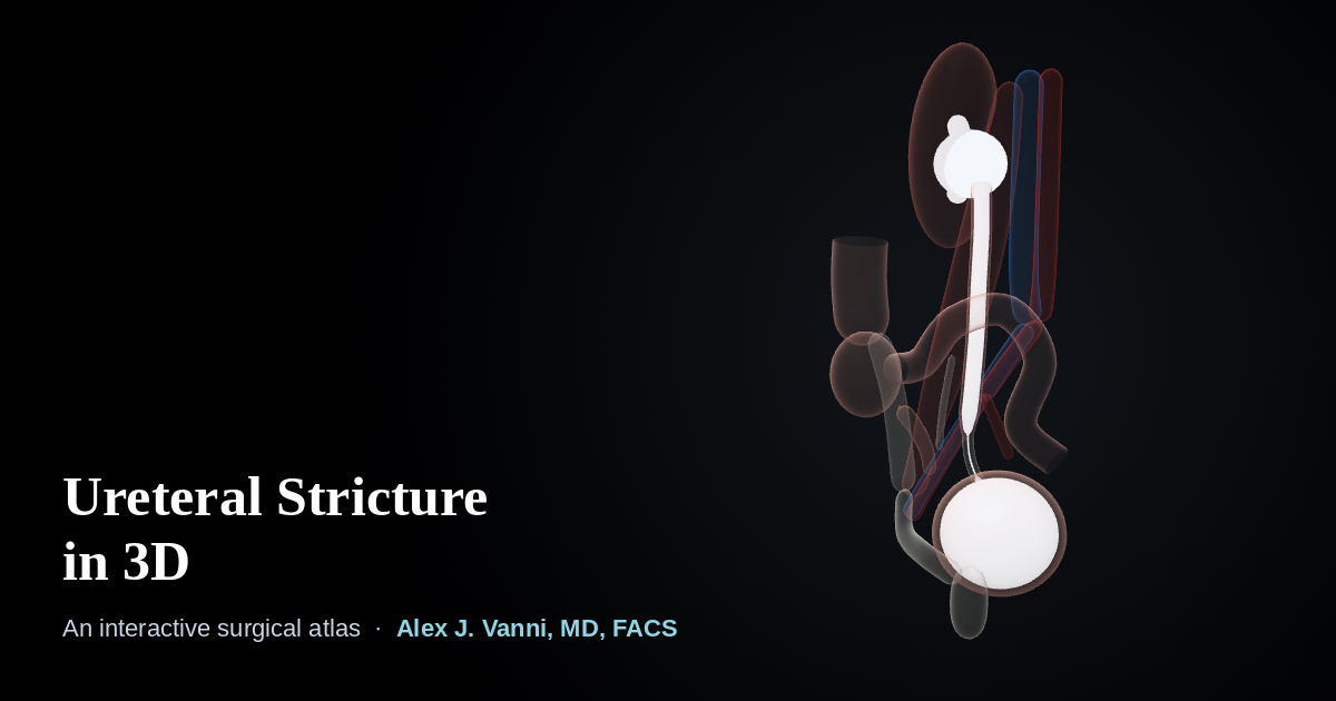
The interactive model needs WebGL, which is not available in this browser. The anatomy and each procedure are described in full below.
Schematic teaching model. Anatomy is simplified and not to scale in every detail. For education only, not for clinical use.
How to Use the Atlas
- Look aroundDrag to rotate. Pinch, or hold Ctrl and scroll, to zoom. Shift-drag moves the model.
- Look insideCutaway opens the model through the ureter. X-ray view shows the collecting system, ureter and bladder the way a contrast study does.
- Ask what something isClick any structure for its name and its part in the repair.
What the Seven Views Show
-
The stricture
A ureteral stricture is scar that narrows the tube carrying urine from the kidney to the bladder. Urine backs up, and the ureter and kidney above it swell (hydronephrosis). The model shows a stricture in the lower (distal) ureter, below where it crosses the iliac vessels.
-
Ureteroureterostomy
For a short stricture, the scarred segment is removed and the two healthy ends are opened slightly and sewn together. Ureteroureterostomy achieves 90 to 95% success for short strictures.
-
Reimplantation with psoas hitch
For a stricture close to the bladder, the ureter is divided above it and reimplanted into the bladder. To bridge the gap without tension, the bladder is stretched up and stitched to the tendon of the psoas muscle.
-
Boari flap
For a longer gap, a flap of bladder wall is cut, rolled into a tube and turned up to reach the ureter, which is then joined to its end. The bladder is usually hitched to the psoas as well.
-
Appendiceal onlay
On the right side, the ureter can be opened along the stricture and the appendix, kept alive on its own blood supply, opened and laid over the gap as a patch.
-
Robotic buccal mucosa graft ureteroplasty
For a stricture in the upper (proximal) ureter, the ureter is opened robotically along the scar and a graft from the lining of the cheek is sewn in as a patch, then wrapped with omentum, a fatty layer with a rich blood supply.
-
Ileal ureter
When the stricture runs the length of the ureter (pan-ureteral), a segment of small intestine (ileum) is isolated on its blood supply and used to replace the ureter, from the kidney to the bladder. The bowel is rejoined behind it.
After most repairs a stent rests inside the ureter while it heals, and it is removed 4 to 6 weeks later in the office. Complex reconstructions achieve 80 to 90% success in experienced hands. See the ureteral stricture FAQs.
The Structures Involved
- Ureter
- The muscular tube, about the width of a pencil lead inside, that carries urine from the kidney to the bladder. It is described in thirds: proximal (upper), mid and distal (lower).
- Iliac vessels
- The large artery and vein to the leg. The ureter crosses in front of them at the rim of the pelvis, a useful landmark between the mid and distal ureter.
- Psoas muscle
- A large muscle along the spine and pelvis. Its tendon is a strong anchor for a psoas hitch.
- Hydronephrosis
- Swelling of the kidney and ureter above a blockage, caused by urine backing up.
About This Model
The atlas is a schematic built for teaching. Shapes and proportions are simplified, only the right side is shown, the strictures are constructed examples rather than real patients, and no two strictures are alike. It is not a substitute for an individual evaluation, which usually includes imaging of the kidney and ureter and often a kidney function scan.
This page is for educational purposes and is not a substitute for an individual medical evaluation. Reviewed by Alex J. Vanni, MD, FACS. Last updated September 19, 2026.
Questions About a Ureteral Stricture?
Dr. Vanni treats complex and recurrent ureteral strictures, including those after radiation or prior surgery, for patients from across New England and nationwide.
Call (781) 744-8762 or learn more about ureteral stricture treatment.