Home › Urinary Fistulas › 3D Surgical Atlas
Rectourethral Fistula in 3D: An Interactive Surgical Atlas
By Alex J. Vanni, MD, FACS — Director, Center for Reconstructive Urologic Surgery, Lahey Clinic
One abnormal channel between the urethra and the rectum, and how it is closed for good. Rotate the model, switch to the X-ray view to see the fistula the way a contrast study shows it, then follow the transperineal repair, including the gracilis muscle flap placed to protect it.
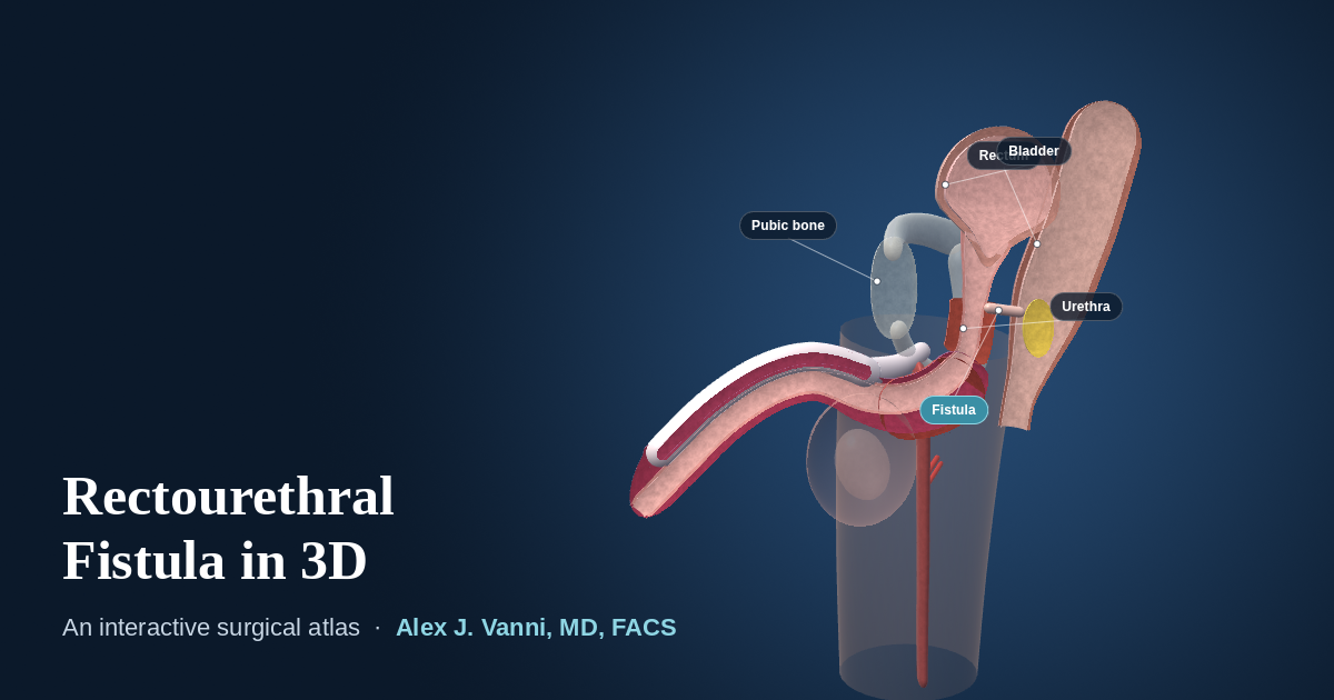
The interactive model needs WebGL, which is not available in this browser. The anatomy and each procedure are described in full below.
Schematic teaching model. Anatomy is simplified and not to scale in every detail. For education only, not for clinical use.
How to Use the Atlas
- Look aroundDrag to rotate. Pinch, or hold Ctrl and scroll, to zoom. Shift-drag moves the model.
- Look insideCutaway opens the model along the midline. X-ray view shows the fistula the way a contrast study does, with contrast passing into the rectum.
- Ask what something isClick any structure for its name and what it has to do with the fistula and its repair.
What the Two Views Show
-
The fistula
A rectourethral fistula is an abnormal channel between the urethra and the rectum. The most common cause is prostate cancer treatment, either surgery or radiation. Urine can pass into the rectum, and gas or stool can pass into the urine, causing infections. On a contrast X-ray, contrast placed in the urethra is seen entering the rectum.
-
Transperineal repair with gracilis flap
Through an incision in the perineum, between the scrotum and the anus, the plane between the rectum and the urethra is opened. The fistula is divided, and the openings in the rectum and in the urethra are each closed separately. In the same operation, the gracilis, a long muscle on the inner thigh that the leg can spare, is freed on its blood supply and placed between the two closures, bringing healthy, well-supplied tissue between them so the channel cannot re-form. This is especially valuable after radiation.
A catheter stays in place for 4 to 6 weeks, and a cystoscopy at 4 to 6 weeks confirms the fistula has closed before it is removed. A temporary colostomy, if needed, is reversed later once the repair has healed. Success is approximately 98% for fistulas after surgery and 86% for radiation-associated fistulas. See recovery after fistula repair and the urinary fistula FAQs.
The Structures Involved
- Urethra
- The channel from the bladder to the tip of the penis. After prostate treatment, a fistula usually opens from the part of the urethra just below the bladder.
- Rectum
- The last part of the bowel, lying directly behind the prostate area and the urethra.
- Perineum
- The area between the scrotum and the anus. The transperineal approach reaches the fistula through it.
- Gracilis muscle
- A long, thin muscle on the inner thigh. Its blood supply enters near the top, so it can be swung up into the perineum without losing it.
About This Model
The atlas is a schematic built for teaching. Shapes and proportions are simplified, the fistula is a constructed example rather than a real patient, and no two fistulas are alike. It is not a substitute for an individual evaluation, which usually includes cystoscopy, contrast imaging and an assessment of the effects of any prior radiation.
This page is for educational purposes and is not a substitute for an individual medical evaluation. Reviewed by Alex J. Vanni, MD, FACS. Last updated September 19, 2026.
Questions About a Rectourethral Fistula?
Dr. Vanni repairs rectourethral fistulas, including radiation-associated and previously repaired fistulas, for patients from across New England and nationwide.
Call (781) 744-8762 or learn more about urinary fistula repair.